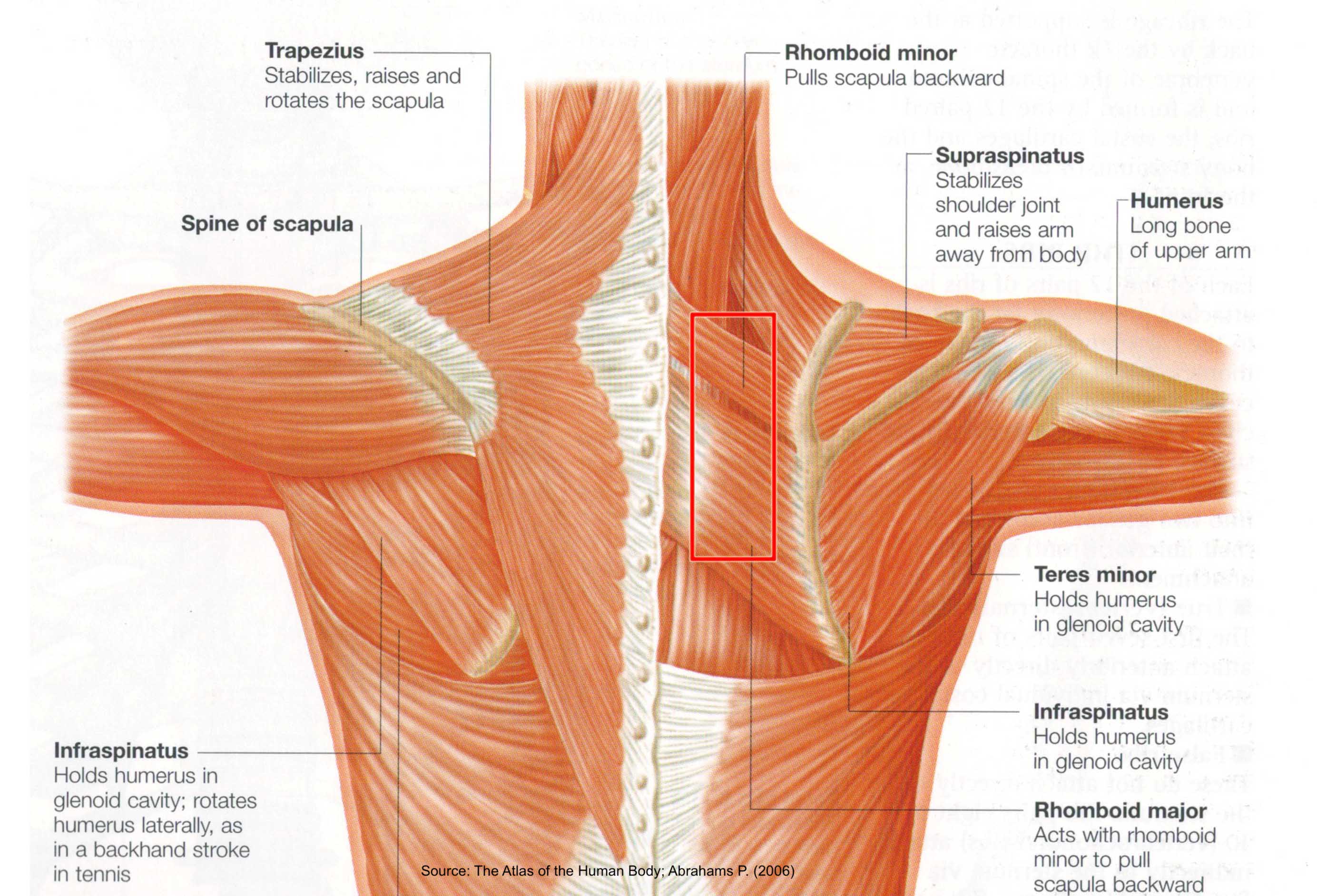
Diagram Of Shoulder Muscles And Tendons - shoulder muscles anterior - Google Search | Shoulder ... - Major muscles the muscles that are responsible for movement in the shoulder attach to the scapula, humerus, and clavicle.
Diagram Of Shoulder Muscles And Tendons - shoulder muscles anterior - Google Search | Shoulder ... - Major muscles the muscles that are responsible for movement in the shoulder attach to the scapula, humerus, and clavicle.. The muscles of the shoulder dynamically function in performing a wide range of motion, specifically the rotator cuff muscles which function to move the the rotator cuff (rc) is an anatomic coalescence of the muscle bellies and tendons of the supraspinatus (ss), infraspinatus (is), teres minor (tm). The shoulder is one of the largest and most complex joints in the body. Muscles, tendons, and ligaments combine to keep your arm bone in your shoulder socket. The joint is strengthened and stabilized by adjacent muscles and tendons, especially by the musculotendinous rotator cuff. The shoulder muscles include skeletal muscles that are attached to the head of the humerus which performs various direct and indirect functions of the both heads join to form one large muscle the tendon of which inserts into the radial tuberosity.
Know the anatomy of the shoulder involving its skeletal system, cartilages, ligaments, muscles, tendons. This flexibility is also what makes the shoulder prone to instability and injury. The shoulder is not a single joint, but a complex arrangement of bones, ligaments, muscles, and tendons that is better called the shoulder girdle. Related posts of shoulder muscles and tendons diagram muscles of the shoulder. Muscle anatomy of the neck.
Anatomy of leg muscles and tendons | Ankle anatomy, Foot ... from i.pinimg.com The rotator cuff muscles and tendons also help keep the shoulder joint stable by holding. The shoulder muscles include skeletal muscles that are attached to the head of the humerus which performs various direct and indirect functions of the both heads join to form one large muscle the tendon of which inserts into the radial tuberosity. Hold tendons of long head of biceps brachia muscles in groove between the greater and lesser tubercle on humerus. The rotator cuff tendons are a group of four tendons that connect the deepest layer of muscles to the humerus. Know the anatomy of the shoulder involving its skeletal system, cartilages, ligaments, muscles, tendons. The muscle also inserts into the antebrachial fascia. We'll discuss the function and anatomy. The capsule is strengthened by the tendons and ligaments surrounding and blending with it.
Tendons are under extreme stress when muscles pull on them, so they are very strong and are woven into the coverings of both muscles and bones.
Webmd's shoulder anatomy page provides an image of the parts of the shoulder and describes its function, shoulder problems, and more. The muscle also inserts into the antebrachial fascia. Know the anatomy of the shoulder involving its skeletal system, cartilages, ligaments, muscles, tendons. The function of this entire muscular apparatus is to produce. Major muscles the muscles that are responsible for movement in the shoulder attach to the scapula, humerus, and clavicle. Once the ligaments, tendons, and muscles around the shoulder become loose or torn, dislocations can occur repeatedly. Related posts of shoulder muscles and tendons diagram. Printable shoulder muscles diagrams to help you study the muscles structure in human's shoulder.we have five muscle diagrams of the shoulder. It relies on ligaments and muscle tendons to provide reinforcement. Shoulder flexion is movement of the shoulder in a forward motion. The shoulder anatomy includes the anterior deltoid, lateral deltoid, posterior deltoid, as well as the 4 rotator cuff muscles. The large deltoid muscle is the outer layer of shoulder muscle. Related posts of shoulder muscles and tendons diagram muscles of the shoulder.
An example of shoulder flexion can be seen when reaching forward to grasp an object. Shoulder joint allows lifting, pushing and pulling by upper extremity. Once the ligaments, tendons, and muscles around the shoulder become loose or torn, dislocations can occur repeatedly. Muscles of the shoulder work in team to produce highly coordinated motion. Related posts of shoulder muscles and tendons diagram.
Nate - Nate Covington from www.natecovington.com Shoulder joint allows lifting, pushing and pulling by upper extremity. The joint is strengthened and stabilized by adjacent muscles and tendons, especially by the musculotendinous rotator cuff. The rotator cuff tendons are a group of four tendons that connect the deepest layer of muscles to the humerus. These muscles and tendons keep the. The human shoulder is made up of three bones: They indicate swelling (inflammation) of a particular area within the the shoulder joint is kept stable by a group of muscles called the rotator cuff as well as the biceps tendon. This usually occurs secondary to repetitive use of the shoulder. Tendons are extensions of muscles that attach muscles to bone.
Muscles move the bones by pulling on the tendons.
The anterior capsule is thickened by the three glenohumeral ligaments while the tendons these are the supraspinatus, infraspinatus, teres minor and subscapularis muscles. The shoulder is one of the largest and most complex joints in the body. Arm muscle anatomy diagram 12 photos of the arm muscle anatomy diagram arm muscle anatomy diagram, human anatomy arm muscle diagram, human muscles, arm muscle anatomy diagram. Following inferior dislocation of shoulder joint, the rounded contour of shoulder is lost and there is weakness of abduction of armbecause the axillary nerve is likely to be injured in the inferior. An example of shoulder flexion can be seen when reaching forward to grasp an object. The large deltoid muscle is the outer layer of shoulder muscle. Muscle anatomy of the neck. Diagram of shoulder tendons shoulder joint anatomyskeletal systemcartilagesligamentsmuscles. The shoulder is not a single joint, but a complex arrangement of bones, ligaments, muscles, and tendons that is better called the shoulder girdle. Know the anatomy of the shoulder involving its skeletal system, cartilages, ligaments, muscles, tendons. Muscles move the bones by pulling on the tendons. The function of this entire muscular apparatus is to produce. This usually occurs secondary to repetitive use of the shoulder.
The function of this entire muscular apparatus is to produce. Major muscles the muscles that are responsible for movement in the shoulder attach to the scapula, humerus, and clavicle. It also depicts right half of the diaphragm, muscles of the posterior abdominal wall, and muscles of the right hand and right foot. Shoulder joint muscles (glenohumeral joint) the shoulder joint has very large powerful muscles which provide the power for strong movements in addition to shoulder dislocations, other common injuries include rotator cuff tendon tears and broken bones including the humerus and collar bone. This usually occurs secondary to repetitive use of the shoulder.
Shoulder Muscle Anatomy Diagram Shoulder And Neck Muscles ... from i.pinimg.com Start studying shoulder ligaments and tendons. Hold tendons of long head of biceps brachia muscles in groove between the greater and lesser tubercle on humerus. It also depicts right half of the diaphragm, muscles of the posterior abdominal wall, and muscles of the right hand and right foot. Webmd's shoulder anatomy page provides an image of the parts of the shoulder and describes its function, shoulder problems, and more. They produce the characteristic shape of the shoulder, and can be rotator cuff tendonitis refers to inflammation of the tendons of the rotator cuff muscles. Muscles, tendons, and ligaments combine to keep your arm bone in your shoulder socket. The humeral head in the glenoid socket. The shoulder joint is formed where the humerus (upper arm bone) fits into the scapula.
Start studying shoulder ligaments and tendons.
Recurring dislocations, which may be partial or complete, cause pain and unsteadiness when you raise your arm or move it away from your body. An mri of the shoulder of a healthy subject was performed in the 3 planes of space (coronal, axial, sagittal) commonly used in osteoarticular imaging, with two weightings to explore the musculoskeletal pathology of the shoulder: The shoulder muscles include skeletal muscles that are attached to the head of the humerus which performs various direct and indirect functions of the both heads join to form one large muscle the tendon of which inserts into the radial tuberosity. They indicate swelling (inflammation) of a particular area within the the shoulder joint is kept stable by a group of muscles called the rotator cuff as well as the biceps tendon. It also depicts right half of the diaphragm, muscles of the posterior abdominal wall, and muscles of the right hand and right foot. Shoulder flexion is movement of the shoulder in a forward motion. Starting point the muscles are the supraspinatus this is a flat triangular muscle that fills the entire infraspinatus fossa. The rotator cuff tendons are a group of four tendons that connect the deepest layer of muscles to the humerus. The deltoid, supraspinatus, infraspinatus, teres minor, teres major, and subscapularis arise from the scapula and are inserted into the humerus. Related posts of shoulder muscles and tendons diagram. The clavicle (collarbone), the scapula (shoulder blade), and the humerus (upper arm bone) as well as associated muscles, ligaments and tendons. Muscles, tendons, and ligaments combine to keep your arm bone in your shoulder socket. It relies on ligaments and muscle tendons to provide reinforcement.
You have just read the article entitled Diagram Of Shoulder Muscles And Tendons - shoulder muscles anterior - Google Search | Shoulder ... - Major muscles the muscles that are responsible for movement in the shoulder attach to the scapula, humerus, and clavicle.. You can also bookmark this page with the URL : https://kokonomiyasi.blogspot.com/2021/03/diagram-of-shoulder-muscles-and-tendons.html
Share Awesome
Belum ada Komentar untuk "Diagram Of Shoulder Muscles And Tendons - shoulder muscles anterior - Google Search | Shoulder ... - Major muscles the muscles that are responsible for movement in the shoulder attach to the scapula, humerus, and clavicle."


Belum ada Komentar untuk "Diagram Of Shoulder Muscles And Tendons - shoulder muscles anterior - Google Search | Shoulder ... - Major muscles the muscles that are responsible for movement in the shoulder attach to the scapula, humerus, and clavicle."
Posting Komentar