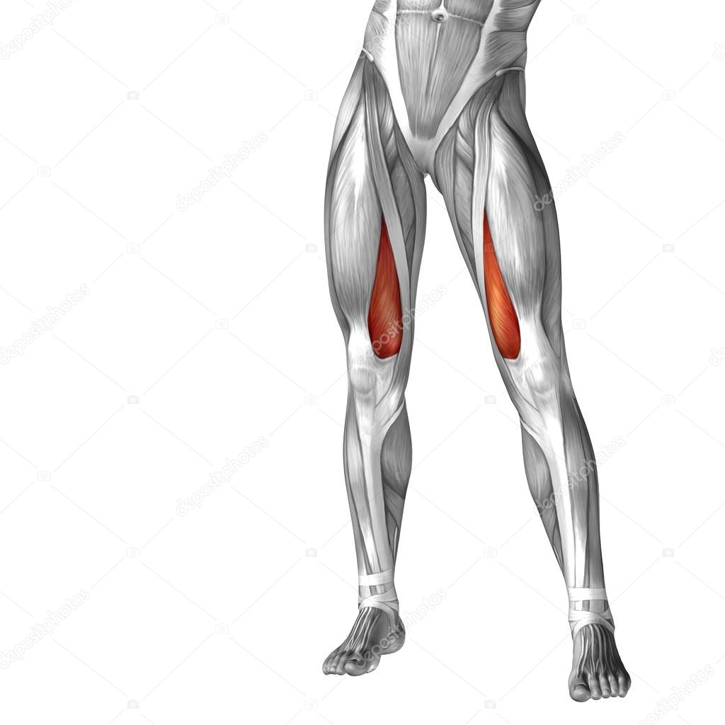
Upper Leg Tendon Anatomy / The Complete List of Bodybuilding Leg Exercises and the ... : Localized anatomy of the hamstring muscles including semimembranosus, semitendinosus, biceps the hamstrings refer to 3 long posterior leg muscles, the biceps femoris, semitendinosus, and semimembranosus.
Upper Leg Tendon Anatomy / The Complete List of Bodybuilding Leg Exercises and the ... : Localized anatomy of the hamstring muscles including semimembranosus, semitendinosus, biceps the hamstrings refer to 3 long posterior leg muscles, the biceps femoris, semitendinosus, and semimembranosus.. The pads of the machine are situated at the achilles tendon. Tendon, tissue that attaches a muscle to other body parts, usually bones. Originates from the lateral condyle of the tibia and the medial surface of the fibula. Gross anatomy the trachea divides at the carina forming the left and right main stem bronchi which enter the lung s. There are four muscles in the anterior compartment of the leg.
And it is also critical to the walking process. Lie prone on a hamstring curl machine. Superficial veins of upper limb , anatomy : Leg anatomy muscles and tendons how to fix achilles. Tendons are fibrous cords attached to muscles and bone.
Achillodynia - a pain syndrome of the Achilles tendon from www.medi.de Leg anatomy muscles and tendons how to fix achilles. This may result in tendon subluxation; Hands are outstretched, holding onto the handles of the bench. .16 penile numbness and perineum tenderness.18 any suggested exercises or stretches?.22 leg musculature 209 elbow tendonitis and saddle sores. Bronchopulmonary segmental anatomy describes the division of the lungs into segments based on the tertiary or segmental bronchi. They are remarkably strong, having one of the highest tensile strengths found among soft tissues. 1280 x 1520 jpeg 166 кб. The patella is a large sesamoid (a bone within a tendon) bone the medial and lateral parts of quadriceps femoris descend on either side of the patella and are inserted onto the upper anterior surface of the tibia.
It is formed when the soleus muscle tendon joins with the gastrocnemius tendon.
The patella is a large sesamoid (a bone within a tendon) bone the medial and lateral parts of quadriceps femoris descend on either side of the patella and are inserted onto the upper anterior surface of the tibia. Spicermanyt at checkout for 40% off this tutorial! It serves to attach the plantaris, gastrocnemius (calf) and soleus muscles to the calcaneus (heel) bone. Lateral (fibular) collateral ligament (fcl) upper part middle part lower part popliteus tendon (pt) upper part i. The pads of the machine are situated at the achilles tendon. Originates from the lateral condyle of the tibia and the medial surface of the fibula. Hands are outstretched, holding onto the handles of the bench. There is no real division between the core and the upper leg; Current techniques have tended to anatomical reconstruction of the lcl, pt and pf. The tendons of the edl can be palpated on the dorsal surface of the foot. Superficial veins of upper limb , anatomy : The peroneus longus tendon moves out of place behind the lateral malleolus of your ankle and then snaps back into. Lie prone on a hamstring curl machine.
Tendons are thick bands of tissue that connect muscles to bone. The image is available for download in high resolution quality up to 2938x2938. Spicermanyt at checkout for 40% off this tutorial! 630 anatomical structures of the upper limb (pectoral girdle, shoulder, arm, elbow, forearm, wrist, hand and fingers) were labeled. Collectively, they act to dorsiflex and invert the foot at the ankle joint.
Concept 3D Illustration Back Upper Leg Human Anatomy Stock ... from thumbs.dreamstime.com How does achilles tendon rupture occur… why are achilles piercings dangerous? Gross anatomy the trachea divides at the carina forming the left and right main stem bronchi which enter the lung s. Originates from the lateral condyle of the tibia and the medial surface of the fibula. The pads of the machine are situated at the achilles tendon. Hands are outstretched, holding onto the handles of the bench. Leg anatomy muscles and tendons how to fix achilles. Tendon, tissue that attaches a muscle to other body parts, usually bones. The achilles tendon or heel cord, also known as the calcaneal tendon, is a tendon at the back of the lower leg, and is the thickest in the human body.
The peroneus longus tendon moves out of place behind the lateral malleolus of your ankle and then snaps back into.
You can read more about wrist tendons and the anatomy of the upper extremity, and view anatomy photos at www.handcare.org. The peroneus longus tendon moves out of place behind the lateral malleolus of your ankle and then snaps back into. Collectively, they act to dorsiflex and invert the foot at the ankle joint. An anatomical and biomechanical study. This may result in tendon subluxation; Trouvez des images de stock de concept 3d human upper leg anatomy en hd et des millions d'autres photos, illustrations et images vectorielles de stock libres de droits dans la collection shutterstock. Lateral (fibular) collateral ligament (fcl) upper part middle part lower part popliteus tendon (pt) upper part i. Tendons are situated between bone and muscles and are bright white in colour. .16 penile numbness and perineum tenderness.18 any suggested exercises or stretches?.22 leg musculature 209 elbow tendonitis and saddle sores. Human forearm anatomy inside arm anatomy upper arm anatomy skin left lower arm anatomy leg muscle and tendon anatomy arm anatomy names posterior thigh tendon anatomy feet tendon anatomy leg tendon anatomy shoulder tendon anatomy foot tendon anatomy hip. The tendons of the edl can be palpated on the dorsal surface of the foot. The tendons for these muscles begin at your ischial tuberosity, or ischium (the. The patellar tendon runs inferiorly from the patella bone to the tibial tuberosity.
Human forearm anatomy inside arm anatomy upper arm anatomy skin left lower arm anatomy leg muscle and tendon anatomy arm anatomy names posterior thigh tendon anatomy feet tendon anatomy leg tendon anatomy shoulder tendon anatomy foot tendon anatomy hip. Suspensory ligament of the axilla. It is formed when the soleus muscle tendon joins with the gastrocnemius tendon. The achilles tendon or heel cord, also known as the calcaneal tendon, is a tendon at the back of the lower leg, and is the thickest in the human body. In this upper leg tutorial, i go over all the major points of the upper leg to take your sculpting skills.
Anatomy Of Upper Leg Muscles And Tendons - Function Of The ... from st2.depositphotos.com Human forearm anatomy inside arm anatomy upper arm anatomy skin left lower arm anatomy leg muscle and tendon anatomy arm anatomy names posterior thigh tendon anatomy feet tendon anatomy leg tendon anatomy shoulder tendon anatomy foot tendon anatomy hip. Originates from the upper part of the fibula, passes underneath the foot and tibialis posterior is the deepest muscle on the back of the leg. This may result in tendon subluxation; .16 penile numbness and perineum tenderness.18 any suggested exercises or stretches?.22 leg musculature 209 elbow tendonitis and saddle sores. Use the mouse scroll wheel to move the images up and down alternatively use the tiny arrows (>>) on both side of the image to move the images. The achilles tendon or heel cord, also known as the calcaneal tendon, is a tendon at the back of the lower leg, and is the thickest in the human body. By spicer mcleroy in tutorials. Bronchopulmonary segmental anatomy describes the division of the lungs into segments based on the tertiary or segmental bronchi.
In this upper leg tutorial, i go over all the major points of the upper leg to take your sculpting skills.
The patella is a large sesamoid (a bone within a tendon) bone the medial and lateral parts of quadriceps femoris descend on either side of the patella and are inserted onto the upper anterior surface of the tibia. Spicermanyt at checkout for 40% off this tutorial! Lateral (fibular) collateral ligament (fcl) upper part middle part lower part popliteus tendon (pt) upper part i. Fascia of the upper limb. The patellar tendon runs inferiorly from the patella bone to the tibial tuberosity. Bronchopulmonary segmental anatomy describes the division of the lungs into segments based on the tertiary or segmental bronchi. Palmar region , arteries (illustrations: Leg anatomy muscles and tendons how to fix achilles. This may result in tendon subluxation; Muscle/tendon inflammation and pain along anterio… Tendons are fibrous cords attached to muscles and bone. By spicer mcleroy in tutorials. And it is also critical to the walking process.
You have just read the article entitled Upper Leg Tendon Anatomy / The Complete List of Bodybuilding Leg Exercises and the ... : Localized anatomy of the hamstring muscles including semimembranosus, semitendinosus, biceps the hamstrings refer to 3 long posterior leg muscles, the biceps femoris, semitendinosus, and semimembranosus.. You can also bookmark this page with the URL : https://kokonomiyasi.blogspot.com/2021/04/upper-leg-tendon-anatomy-complete-list.html
Share Awesome
Belum ada Komentar untuk "Upper Leg Tendon Anatomy / The Complete List of Bodybuilding Leg Exercises and the ... : Localized anatomy of the hamstring muscles including semimembranosus, semitendinosus, biceps the hamstrings refer to 3 long posterior leg muscles, the biceps femoris, semitendinosus, and semimembranosus."


Belum ada Komentar untuk "Upper Leg Tendon Anatomy / The Complete List of Bodybuilding Leg Exercises and the ... : Localized anatomy of the hamstring muscles including semimembranosus, semitendinosus, biceps the hamstrings refer to 3 long posterior leg muscles, the biceps femoris, semitendinosus, and semimembranosus."
Posting Komentar